 MENUBEFORE & AFTERPATIENT REVIEWSMENUBEFORE & AFTERPATIENT REVIEWS
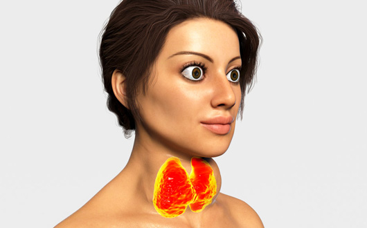
Graves’ disease is an autoimmune condition that causes the thyroid gland to become overactive (hyperthyroidism). In this disease, the immune system mistakenly attacks the body’s own tissues, affecting not only the thyroid gland but also the muscles and fat around the eyes. This condition is known as “Thyroid Eye Disease” or “Graves’ Ophthalmopathy.”
If left untreated, it can lead to permanent eye damage and vision loss. The most common symptoms include:
As the immune system attacks the thyroid gland, it also targets the tissues around the eyes due to their similar structure. This attack causes inflammation and enlargement of the muscles and fat behind the eyes. As a result, the eyes are pushed forward, the eyelids widen, and the surface of the eyes can be damaged.
Graves’ disease can occur at any age and in both sexes, but it is most commonly seen in women between the ages of 30 and 60. Factors that increase the risk of developing the disease include:
The primary cause of thyroid eye disease is a malfunction in the immune system. Normally, the immune system does not attack the body’s own tissues; however, in Graves’ disease, antibodies called TSH receptor antibodies bind to the thyroid gland and the tissues around the eyes.
This process leads to excessive hormone production and enlargement of the thyroid gland, while also causing swelling and inflammation in the muscles and fat tissue around the eyes. As a result, symptoms such as eye protrusion, swelling, redness, and restricted eye movement may appear. The severity of the disease varies from person to person and is directly related to the intensity of the immune response.
Most people with Graves’ disease have hyperthyroidism, but in some cases, thyroid hormone levels may be normal or even low. Sometimes, eye symptoms appear months before thyroid abnormalities, serving as the first warning sign. In some patients, eye symptoms become more pronounced during the transition from a hypothyroid to a hyperthyroid state. Rarely, eye signs may appear at the time of diagnosis of a thyroid nodule or thyroid cancer.
The symptoms of thyroid eye disease vary between individuals and can range from mild to severe. You should consult an oculoplastic surgeon if you experience any of the following:
Graves’ disease can follow a different course in each patient. In some cases, the condition may improve spontaneously, while in others, it may progress severely and lead to vision loss. The disease generally occurs in two phases: an active (inflammatory) phase and an inactive (fibrotic) phase.
During the active phase, eye redness, swelling, and discomfort are prominent. In the inactive phase, inflammation decreases, but eye protrusion and eyelid abnormalities may become permanent. The disease usually stabilizes within 1–2 years, although this period can vary between individuals. Thyroid hormone levels do not always correlate directly with the severity of eye symptoms.
Diagnosis of thyroid eye disease requires a collaborative approach between endocrinologists and oculoplastic surgeons. The evaluations performed for diagnosis include:
Treatment of Graves’ disease is planned according to the disease phase. The goal is to control inflammation during the active phase and correct permanent deformities during the inactive phase.
Once inflammation subsides, surgical treatment may be applied for permanent deformities:
The typical surgical sequence is: decompression → strabismus surgery → eyelid surgery. In some patients, two procedures can be performed simultaneously.
Orbital decompression surgery is performed to reduce eye protrusion (proptosis) caused by thyroid eye disease. Inflammation causes thickening of the orbital muscles and fat, which pushes the eyeball forward. This can create an undesirable cosmetic appearance and may compress the optic nerve, leading to vision loss.
The aim of the surgery is to reduce pressure behind the eye and allow it to return to a more natural position. This is achieved by carefully expanding one or more of the orbital bone walls, creating additional space for the tissues behind the eye.
Orbital decompression surgery is indicated in the following situations:
How Is Orbital Decompression Surgery Performed?
Orbital decompression is a surgical procedure performed under general anesthesia. The surgery is usually carried out through the inside of the eyelid (transconjunctival) or via hidden skin incisions.
Depending on the severity of the disease, one, two, or three orbital walls may be decompressed. In medial (inner wall) decompression, the bone on the inner side of the eye is thinned to reduce pressure. Inferior (lower wall) decompression relieves the eye downward and inward, while lateral (outer wall) decompression provides the most noticeable cosmetic improvement. In some cases, fat decompression can be performed alone or in combination with bone decompression. In mild cases, fat decompression alone may be sufficient.
Postoperatively, patients are usually discharged on the same day or the next day. Recovery generally takes 1–2 weeks, although complete tissue stabilization may require 3–6 months. Temporary bruising, swelling, or double vision may occur. During the first two weeks, patients are advised to avoid sneezing and strenuous activities.
Benefits of Orbital Decompression Surgery
Orbital decompression significantly reduces eye protrusion caused by thyroid eye disease. It relieves pressure on the optic nerve, corrects eyelid position, and reduces dryness and discomfort. Additionally, by restoring balance to the tissues around the eyes, the surgery improves facial symmetry and overall aesthetic appearance.
Risks of Orbital Decompression Surgery
As with any surgical procedure, orbital decompression carries certain risks. These may include temporary or permanent double vision, changes in eyelid position, and, rarely, infection or bleeding. Mild asymmetry may require additional corrective procedures. Therefore, it is essential that the surgery is performed by an experienced oculoplastic surgeon.
Surgery for thyroid eye disease may be urgently required in certain situations. These include optic nerve compression (optic neuropathy),rapidly increasing eye protrusion, and the risk of corneal erosion or perforation due to severe dryness. In other cases, surgery is planned after the disease has remained stable for at least six months. Additionally, it is important that TSH receptor antibody levels have normalized and that the patient has stopped smoking before undergoing surgery.
Thyroid eye disease can be distressing not only medically but also cosmetically, often creating a tired, surprised, or angry facial expression. Modern oculoplastic surgery can restore both functional and aesthetic balance, providing a natural and refreshed appearance. Surgical procedures can correct eye protrusion, eyelid position, enlarged fat tissue, and the shape of the lacrimal gland.
The cost of thyroid eye disease surgery varies depending on the stage of the disease, the type of surgery performed, whether it is unilateral or bilateral, and the surgeon’s experience. Because the treatment plan is individualized for each patient, the exact cost can only be determined after an in-person consultation.


 Altuğ Çetinkaya, MD, FEBOOphthalmologist Turkey