 MENUBEFORE & AFTERPATIENT REVIEWSMENUBEFORE & AFTERPATIENT REVIEWS
With over 25 years of experience in the field of Oculoplastic Surgery, both internationally and domestically, I accept patients at my private clinic located in Ankara Maidan Business Center. In line with the principles of patient health and honest medical practice, all surgical and non-surgical, cosmetic and functional procedures related to Oculoplastic and Orbital Surgery being performed worldwide can be carried out at our clinic.
 Blepharoplasty Turkey (Aesthetic Eyelid Surgery)
 Upper Eyelid Blepharoplasty in Turkey
 Lower Blepharoplasty in Turkey
 Revision Eyelid Surgery (Revision Blepharoplasty)
 Drooping Eyelids (Ptosis) Turkey
 Eyebrow Lift Surgery Turkey
 Blepharospasm (Eyelid Spasm)
 Non-Surgical Eye Area Aesthetics
 Eyelid Tumors
 Blocked Tear Duct Treatment Turkey
 Watery Eye Treatment Turkey
 Orbital Tumors
Your experience begins with direct and discreet contact via WhatsApp. You may initially share photographs and brief information regarding your concerns, allowing us to gain a preliminary understanding and begin a personalized dialogue.
While this is a helpful first step, true excellence in eyelid and oculoplastic surgery cannot be achieved through photographs alone. These procedures require meticulous evaluation, precise measurements, and a comprehensive understanding of facial dynamics. For this reason, treatment decisions and exact pricing are never finalized based solely on images, as doing so may compromise accuracy and long-term outcomes.
You may schedule a private online consultation at your convenience. During this one-on-one video meeting, Dr. Çetinkaya evaluates your condition in detail, discusses appropriate treatment options, and answers your questions thoroughly.
At this stage, based on a professional clinical assessment, a preliminary (ballpark) cost estimate may be provided to assist with planning. This estimate is indicative and may be refined following in-person evaluation.
If you decide to proceed, our team will coordinate your visit with care and precision. Airport transfer arrangements can be provided to ensure a seamless arrival. A comprehensive face-to-face examination is then performed, allowing the treatment plan and associated costs to be confirmed or refined as necessary.
We assist our international patients in arranging high-quality, cost-effective luxury or comfort accommodations conveniently located near the clinic and surgical facilities—ensuring privacy, ease of access, and a calm recovery environment.
To begin your personalized journey or to receive further information, please contact our clinic via WhatsApp: +90 530 279 0315. From your very first message, our team is committed to delivering a bespoke, discreet, and professionally guided experience—centered entirely on your safety, comfort, and confidence.


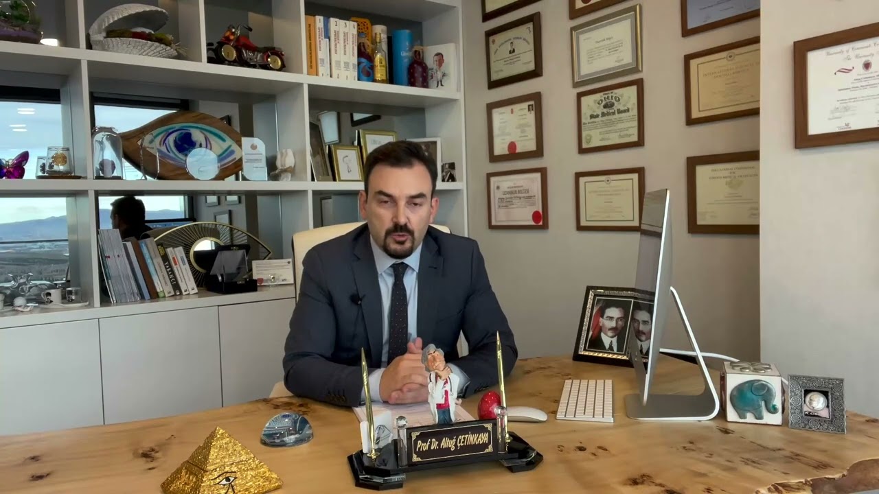
Prof. Dr. Altuğ Çetinkaya has completed a fellowship in Oculoplastic, Orbital, and Reconstructive Surgery in the United States, focusing on correcting cosmetic and structural disorders of the eyelids and surrounding ocular tissues. Oculoplastic surgeons are ophthalmologists who receive advanced training during and after their four-year ophthalmology residency in the diagnosis and surgical treatment of conditions affecting the tissues surrounding the eyes. The primary areas of this specialty include eyelid ptosis/bagging, eyelid deformities, brow ptosis, eyelid and orbital tumors, tear duct obstructions, orbital (eye socket) diseases, thyroid-related eye deformities, periocular trauma, and prosthetic eye treatments.


 Altuğ Çetinkaya, MD, FEBOOphthalmologist Turkey
I am so grateful to have met Prof. Dr. Altuğ Çetinkaya He corrected my previously unsuccessful blepharoplasty, which had been performed by another surgeon, at a time when I had completely lost hope. He carefully and delicately
14.06.2026Read MoreWe had been searching for a good doctor for our son’s under-eye bags for about a year until we met Prof. Dr. Altuğ Çetinkaya. He successfully performed surgery to remove my son’s under-eye bags. I can confidently say he is on
10.06.2026Read MoreI had a blockage in my tear duct and was initially afraid of undergoing surgery. However, Dr. Prof. Dr. Altuğ Çetinkaya was so professional and confident that I decided to proceed without hesitation. He successfully performed
04.06.2026Read MoreWe consulted Prof. Dr. Altuğ Çetinkaya for our 4-year-old child who had congenital ptosis in one eyelid. We had the surgery about two months ago. He is a very experienced doctor who personally took great care of us throughout
01.06.2026Read MoreHello, I am very glad to have met Prof. Dr. Altuğ Çetinkaya. Thank you for your skill and care. My under-eye bags and drooping eyelids were making me feel tired and unwell. However, the idea of surgery was initially frighteni
26.05.2026Read MoreDear Doctor, After extensive research from Germany, I reached you—and I am truly glad I did. Your precise diagnoses and deep expertise enlightened me greatly. Your consistent professionalism and honesty in recommending only wh
17.05.2026Read More